
About

My scientific journey began in Robert Cerny's Lab at Charles University in Prague, where I was first introduced to the captivating realms of embryology and evo-devo. While pursuing my Ph.D., I participated in the 2017 Embryology Course at the Marine Biological Laboratory in Woods Hole, Massachusetts. This experience deepened my passion for studying the intriguing and diverse organisms that play crucial roles in expanding our understanding of evolution and development. My PhD thesis focused on cranial neural crest migration and differentiation in several fishes, with an emphasis on basal lineages such as bichirs and sturgeons. For my postdoctoral studies, I aimed to gain a deeper understanding of the neural crest at the molecular level. To this end, I joined the laboratory of Marianne Bronner at the California Institute of Technology (Caltech) to investigate the role of the neural crest in both development and regeneration in non-model organisms such as lamprey and sturgeon, supported by a Marie Sklodowska-Curie Postdoctoral Fellowship funded by the European Union (co-hosted by Caltech in the United States and the University of South Bohemia in the Czech Republic). My current research is supported by an NIH K99/R00 Pathway to Independence Award, with the goal of elucidating the molecular mechanisms that drive neural crest stem cells to differentiate into dermal bones.
Positions and Professional Stays
NIH NIDCR K99/R00 Fellow, Bronner Lab – Caltech, USA (2025 - Present)
Marie Sklodowska-Curie Postdoctoral Fellow, Bronner Lab & Psenicka Lab - Caltech, USA and University of South Bohemia, Czech Republic (2021-2023)
Postdoctoral Scholar, Bronner Lab – Caltech, USA (2019 - Present)
Guest PhD student, Haitina Lab – Uppsala University, Sweden (2017)
Guest PhD student, Olsson Lab – Friedrich-Schiller University, Germany (2015-2016)
PhD in Zoology, Cerny Lab – Charles University, Czech Republic (2013-2019)
Researcher, Natural History Museum – National Museum, Czech Republic (2012-2019)


Research
Development and evolution of the neural crest odontoskeletogenic ability

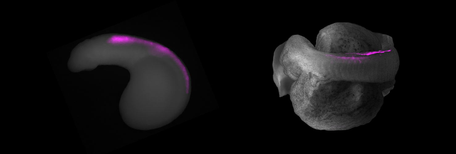
The neural crest is an important stem cell population in the evolution and development of vertebrates, playing a key role in evolving predatory skills and expanding the skull and brain. In vertebrate embryos, the neural crest divides into several subpopulations based on origins, such as the cranial, vagal, and trunk neural crest. Although all these populations share a common set of genes, they are also unique depending on their specific region of origin along the body axis. I use comparative molecular analyses and cell lineage analyses to investigate the developmental potential of neural crest cells across early branching lineages of vertebrates, such as lampreys and sturgeons. My particular focus is on their odontoskeletogenic ability to produce hard tissues such as bone and dentin. The results from these analyses will provide a comprehensive understanding of the molecular mechanisms and alterations within specific developmental programs along the body axis, emphasizing what confers the ability to make bone or dentin.
Selected Publications
Ancient vertebrate dermal armor evolved from trunk neural crest. Stundl J, Martik ML, … & Bronner ME. (2023). The Proceedings of the National Academy of Sciences (PNAS), 120: e2221120120.
neural crest and its role in regeneration

The capacity for scarless regeneration seems to have gradually diminished throughout vertebrate evolution as tissues and organs increased their complexity. Thus, 'basal' vertebrates serve as valuable model systems for gaining deeper insights into tissue repair abilities, as they may unveil ancestral regeneration mechanisms. To study this, I employ a comparative approach, focusing on lampreys and sturgeons, both renowned for their exceptional regenerative potential and important phylogenetic position. Regeneration is a multifaceted process involving various cell populations, with glial cells emerging as particularly significant players. Notably, neural crest cells form all of the glia in the vertebrate peripheral nervous system, raising the possibility that they may represent a stem cell-like population that can repair vertebrate tissues. Thus, my research aims to investigate the role of neural crest stem cells in promoting tissue repair (e.g. heart tissues - supported by MSCA No. 897949) in vertebrates.
Selected Publications
Neural crest gene regulatory networks as drivers of development, diversification and disease. Stundl J, Desingu Rajan AR & Bronner ME (2026). Nature Reviews Molecular Cell Biology, 27,
RECONSTRUCTION OF THE DEVELOPMENTAL CHANGES UNDERLYING THE EARLIEST EVENTS IN VERTEBRATE EVOLUTION

To comprehend the developmental programs driving evolution of the early vertebrate body plan, I utilize a comparative approach, studying a diverse range of phylogenetically important non-model organisms such as lamprey and sturgeon alongside established models like zebrafish and salamanders. I primarily aim to decipher the developmental roles of neural crest and mesodermal cells in the formation of vertebrate skeleton and larval organs.
Selected Publications
Acquisiton of neural crest promoted thyroid evolution from chordate endostyle. Stundl J, Desingu Rajan AR, … & Bronner ME. (2025). Science Advances, 11(32):eadv2657
Neural crest origin of sympathetic neurons at the dawn of vertebrates. Edens BM, Stundl J, Urrutia HA, Bronner, ME. (2024). Nature, 626: 121–124.
Bichir external gills arise via heterochronic shift that accelerates hyoid arch development. Stundl J, Pospisilova A, ... & Cerny R. (2019). eLife, 8: e43531.

PUBLICaTIONS
This research received public funding. If you encounter any difficulties accessing the articles listed below, please feel free to contact me.
ACQUISITION OF NEURAL CREST PROMOTED THYROID EVOLUTION FROM CHORDATE ENDOSTYLE. Stundl J*, Desingu Rajan AR, … & Bronner ME*. Sci Adv. (2025):11(32):eadv2657. doi: 10.1126/sciadv.adv2657. *co-corresponding authors
DENTAL DEVELOPMENT IN THE TROPICAL GAR (ATRACTOSTEUS TROPICUS) AND THE EVOLUTION OF VERTEBRATE DENTITIONS. Pospisilova A, Stundl J, … & Soukup V. Dev Dyn. (2025): Jun 19. doi: 10.1002/dvdy.70055.
TEMPTING FATE: NEURAL CREST INDUCTION ALONG THE BODY AXIS. Stundl J & Bronner ME. Cells Dev. (2025):204000. doi: 10.1016/j.cdev.2025.204000.
GENERATION OF A ZEBRAFISH NEUROFIBROMATOSIS MODEL VIA INDUCIBLE KNOCKOUT OF NF2. Desingu Rajan AR, Huang Y^, Stundl J^,… & Bronner ME. Dis Model Mech. (2024): 17(12):dmm050862. doi: 10.1242/dmm.050862. ^equal contribution
TLX3 MEDIATES NEURONAL DIFFERENTIATION AND PROPER CONDENSATION OF THE DEVELOPING TRIGEMINAL GANGLION. Urrutia HA, Stundl J, Bronner ME. Dev Biol. (2024): 515:79-91. doi: 10.1016/j.ydbio.2024.07.005.
NEURAL CREST ORIGIN OF SYMPATHETIC NEURONS AT THE DAWN OF VERTEBRATES. Edens BM, Stundl J, Urrutia HA, Bronner ME. Nature. (2024): 629(8010):121-126. doi: 10.1038/s41586-024-07297-0.
A LAMPREY NEURAL CELL TYPE ATLAS ILLUMINATES THE ORIGINS OF THE VERTEBRATE BRAIN. Lamanna F, Hervas-Sotomayor F, Oel AP, Jandzik D, Sobrido-Cameán D, Santos-Durán GN, Martik ML, Stundl J, … & Kaessmann H. Nat Ecol Evol. 2023 Oct;7(10):1714-1728. doi: 10.1038/s41559-023-02170-1. Epub 2023 Sep 14. Erratum in: Nat Ecol Evol. (2023): 7(10):1741. doi: 10.1038/s41559-023-02227-1.
PRE-MANDIBULAR PHARYNGEAL POUCHES IN EARLY NON-TELEOST FISH EMBRYOS. Horackova A, Pospisilova A, Stundl J, … & Cerny R. Proc Biol Sci. (2023): 290(2006):20231158. doi: 10.1098/rspb.2023.1158.
ANCIENT VERTEBRATE DERMAL ARMOR EVOLVED FROM TRUNK NEURAL CREST. Stundl J*, Martik ML, Chen D, Raja DA, … & Bronner ME*. Proc Natl Acad Sci U S A. (2023): 120(30):e2221120120. doi: 10.1073/pnas.2221120120. *co-corresponding authors
- > Commentary for PNAS written by Andrew Gillis
A MEDIAN FIN DERIVED FROM THE LATERAL PLATE MESODERM AND THE ORIGIN OF PAIRED FINS. Tzung KW, Lalonde RL, Prummel KD, Mahabaleshwar H, Moran HR, Stundl J,… & Carney TJ. Nature. (2023):618(7965):543-549. doi: 10.1038/s41586-023-06100-w.
EVOLUTION OF THE NITRIC OXIDE SYNTHASE FAMILY IN VERTEBRATES AND NOVEL INSIGHTS IN GILL DEVELOPMENT. Annona G, Sato I, Pascual-Anaya J, Osca D, Braasch I, Voss R, Stundl J, … & D'Aniello S. Proc Biol Sci. (2022):289(1980):20220667. doi: 10.1098/rspb.2022.0667
THE REMARKABLE DYNAMICS IN THE ESTABLISHMENT, REARRANGEMENT, AND LOSS OF DENTITION DURING THE ONTOGENY OF THE STERLET STURGEON. Pospisilova A, Stundl J, … & Soukup V. Dev Dyn. (2022):251(5):826-845. doi: 10.1002/dvdy.444.
EFFICIENT CRISPR MUTAGENESIS IN STURGEON DEMONSTRATES ITS UTILITY IN LARGE, SLOW-MATURING VERTEBRATES. Stundl J, Soukup V, … & Jandzik D. Front Cell Dev Biol. (2022):10:750833. doi: 10.3389/fcell.2022.750833.
EVOLUTION OF NEW CELL TYPES AT THE LATERAL NEURAL BORDER. Stundl J, Bertucci PY, … Bronner ME. Curr Top Dev Biol. (2021):141:173-205. doi: 10.1016/bs.ctdb.2020.11.005.
MIGRATORY PATTERNS AND EVOLUTIONARY PLASTICITY OF CRANIAL NEURAL CREST CELLS IN RAY-FINNED FISHES. Stundl J*, Pospisilova A, … & Cerny R.* Dev Biol. (2020):467(1-2):14-29. doi: 10.1016/j.ydbio.2020.08.007. *co-corresponding authors
EMBRYONIC AND LARVAL DEVELOPMENT OF THE NORTHERN PIKE: AN EMERGING FISH MODEL SYSTEM FOR EVO‐DEVO RESEARCH. Pospisilova A, Brejcha J, … & Stundl J* J Morphol. (2019):280(8):1118-1140. doi: 10.1002/jmor.21005. *corresponding author
BICHIR EXTERNAL GILLS ARISE VIA HETEROCHRONIC SHIFT THAT ACCELERATES HYOID ARCH DEVELOPMENT. Stundl J, Pospisilova A, … & Cerny R. Elife. (2019):8:e43531. doi: 10.7554/eLife.43531.
PRE-ORAL GUT CONTRIBUTES TO FACIAL STRUCTURES IN NON-TELEOST FISHES. Minarik M, Stundl J, … & Cerny R. Nature. (2017):547(7662):209-212. doi: 10.1038/nature23008.

NEWS
Exciting NEWS! My research has been awarded by the NIH NIDCR K99/R00 Pathway to Independence Award (2025).


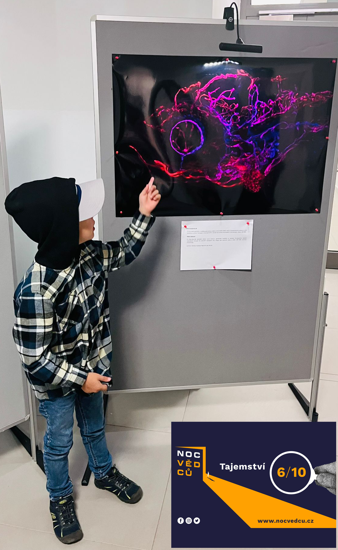
My Research Photographs Featured at European Researchers' Night 2023: 'Mystery' (Tajemstvi Ryb)
I gave an oral presentation at the 20th International Congress of Developmental Biology (co-hosted by SDB & LASDB) in San Juan, Puerto Rico, June 18–22, 2025.


Contact ME
Jan Stundl, Ph.D.
Senior Postdoctoral Scholar Research Associate in Biology and Biological Engineering
NIH NIDCR K99/R00 Fellow
California Institute of Technology
Pasadena, California 91125
><{{{(*> ><{{{(*> ><{{{(*>
